Flavonoid Extract from Propolis Provides Cardioprotection following Myocardial Infarction by Activating PPAR- γ
Evid Based Complement Alternat Med. 2022 Jul 5;2022:1333545
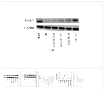
We have previously reported that flavonoid extract from propolis (FP) can improve cardiac function in rats following myocardial infarction (MI). However, the mechanisms responsible for the cardioprotective effects of FP have not been fully elucidated. In the current study, we explored whether FP can reduce inflammatory cytokines and attenuate sympathetic nerve system activity and antiendoplasmic reticulum (ER) stress and whether the cardioprotective effects are related to peroxisome proliferator-activated receptor gamma (PPAR-γ) activation.
Sprague Dawley rats were randomly divided into six groups: Sham group received the surgical procedure but no artery was ligated; MI group received ligation of the left anterior descending (LAD) branch of the coronary artery; MI + FP group received FP (12.5 mg/kg/d, intragastrically) seven days prior to LAD ligation; FP group (Sham group + 12.5 mg/kg/d, intragastrically); MI + FP + GW9662 group received FP prior to LAD ligation with the addition of a specific PPAR-γ inhibitor (GW9662), 1 mg/kg/d, orally); and MI + GW9662 group received the PPAR-γ inhibitor and LAD ligation.
The results demonstrated that the following inflammatory markers were significantly elevated following MI as compared with expression in sham animals: IL-1β, TNF-α, CRP; markers of sympathetic activation: plasma norepinephrine, epinephrine and GAP43, nerve growth factor, thyroid hormone; and ER stress response markers GRP78 and CHOP. Notably, the above changes were attenuated by FP, and GW9662 was able to alleviate the effect of FP.
In conclusion, FP induces a cardioprotective effect following myocardial infarction by activating PPAR-γ, leading to less inflammation, cardiac sympathetic activity, and ER stress.

